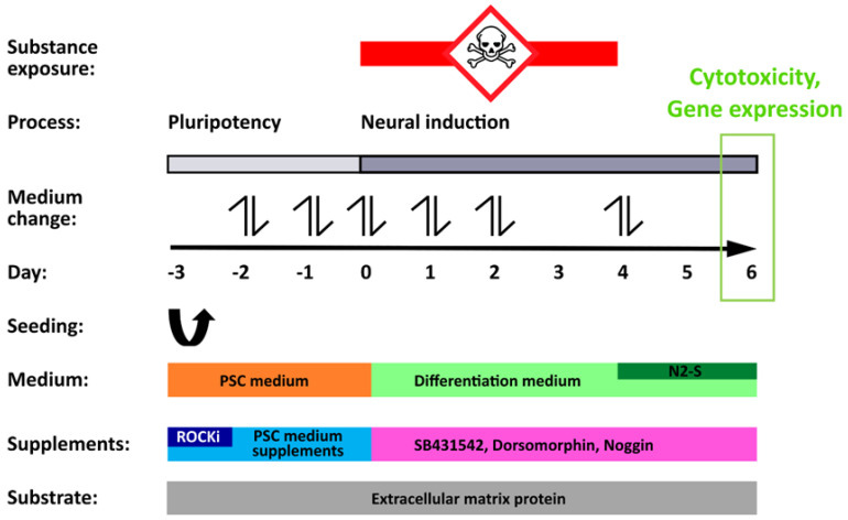
Figure 1.
Schematic representation of the UKN1-test (modified from [20,37]). The overview scheme depicts the differentiation protocol, important experimental steps, and the principal of the toxicity assay. In the pluripotency phase (day −3 to 0), hiPSCs were cultured in a pluripotent stem cell (PSC) medium to maintain their pluripotent state. Factors that inhibited Rho-kinase (ROCKi) were additionally given on the day of seeding (day −3) to support the survival of hiPSCs seeded as single cells on extracellular matrix proteins. From day 0 onwards, the change to a differentiation medium spiked with SB431542, dorsomorphin, and noggin initiated neuroectodermal differentiation of the cells. Simultaneously, cells were exposed to test compounds for a total of 96 h. On day 4, substances were withdrawn and addition of 25% N2-S further enhanced the neural differentiation process. On day 6, compound-induced cytotoxicity was determined and the cells were harvested for gene array analysis. Media changes were conducted as indicated (double arrows) on the days −2, −1, 0, 1, 2, and 4.

