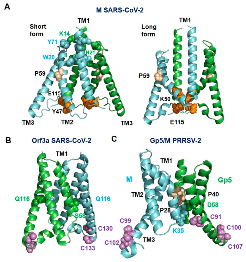
Figure 8.
Structure of the transmembrane region of the short and long form of M (A), Orf3a (B) of SARS-CoV-2 and Gp5/M of PRRSV-2 (C). (A). One monomer of Orf3a and M of SARS-CoV-2 is colored green, and the other is colored cyan. Gp5 is colored green and M is colored cyan in the predicted structure of Gp5/M of the PRRSV-2 reference strain VR 2332. Proline residues in TM2 of M of SARS-CoV-2 and TM1 of Gp5/M are highlighted as wheat spheres, and cysteine residues at the C-terminus of TM3 of Gp5/M and Orf3a are highlighted as magenta spheres. Residues that form hydrophilic interactions between the monomers are highlighted as spheres with the color of the respective monomer. Residues forming interactions between the hinge region and TM2 in the short and long form of M of SARS-CoV-2 are highlighted as orange spheres. Figures were created with Pymol from PDB-file 7VGS (M, short form), 7VGR (M. long form) and 6XDC (Orf3a).

