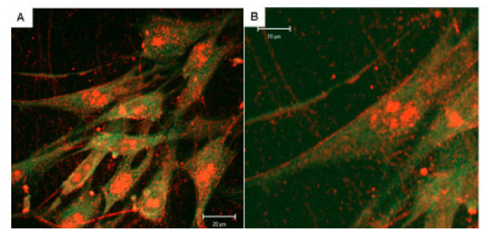
Figure 3.

Fluorescent immunostaining of ARSB in human cerebrovascular cells. (A,B). Confocal microscopy demonstrates cell surface localization of ARSB in untreated cerebrovascular cells, as well as cytoplasmic and nuclear localization and presence of ARSB in cell projections. ARSB is stained red, and β-actin is stained green [44].
