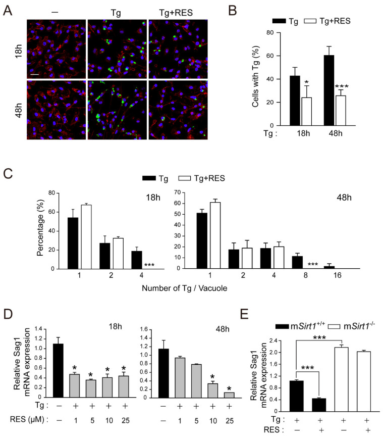
Figure 2.
RES treatment promotes the elimination of intracellular T. gondii in primary murine macrophages. (A–C) BMDMs were infected with a GFP-conjugated RH strain (MOI = 1) for 2 h, followed by the further treatment of RES (10 µM) for the indicated times. Cells were stained with Texas Red®-X phalloidin for F-actin in the cytoskeleton (red) and DAPI for nuclei (blue), respectively. (A) Fluorescent images showing the number of intracellular T. gondii. (B,C) Number of T. gondii RH-infected cells (for B) and T. gondii RH per vacuole were quantified. Scale bar = 25 µm (D,E) qPCR analysis for evaluating Sag1 mRNA expression. (D) T. gondii-infected BMDMs were stimulated with increasing concentrations of RES (1, 5, 10, or 25 µM) for 18 h (for left) or 48 h (for right). (E) BMDMs isolated from mSirt1+/+ and mSirt1−/− mice were infected with RH strain of T. gondii (MOI = 1) for 2 h and then further stimulated with RES (10 µM) for 48 h. Data are representative of three independent experiments and are presented as means ± SD. * p < 0.05 and *** p < 0.001, compared with control cells (two-tailed Student’s t-test). Tg, Toxoplasma gondii; RES, resveratrol.

