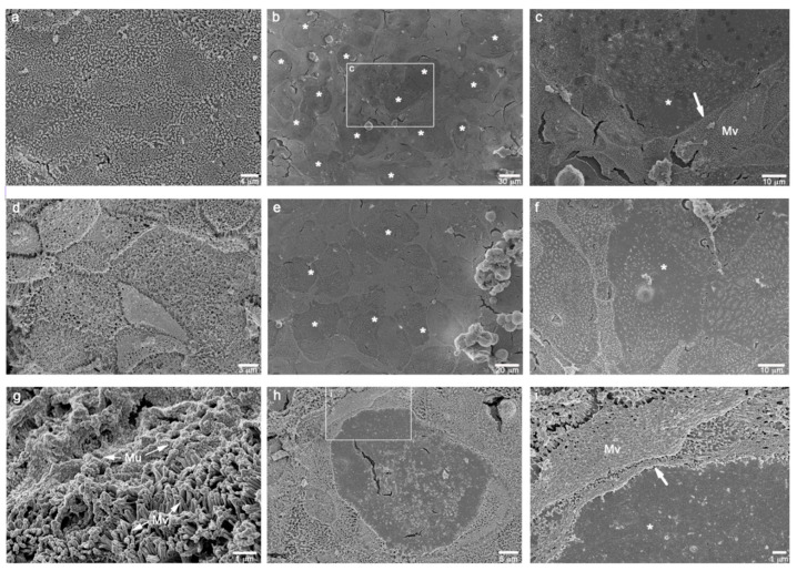
Figure 3.
SEM micrographs of the different combinations of cell cultures grown on inserts, at 21st day of culture. (a): Caco-2 monoculture showing a homogeneous leaflet of polarized epithelial cells with a great abundance of surface microvilli. (b): Caco-2/Raji B co-culture displaying some rounded areas of microvilli loss. (c): high magnification of the squared area in b showing the boundary (white arrow) between presence (Mv) and absence (*) of the surface microvilli. (d): Caco-2/HT29-MTX differentiated co-culture rich in surface microvilli. (e,f): Caco-2/HT29-MTX/Raji B tri-culture showing a lot of inducted smooth rounded areas of possible M-cell phenotype (*). (g): Caco-2/HT29-MTX co-culture displaying a consistent mucus layer (Mu) on the microvilli. (h,i): high magnifications of a feasible M-cell phenotype (*) in Caco-2/HT29-MTX/Raji B tri-culture (white arrow: boundary of the microvilli loss.

