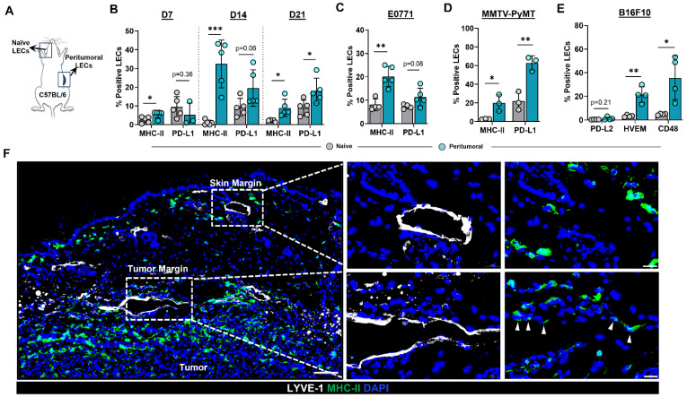
Figure 1.
Peritumoral LECs upregulate MHC-II, PD-L1, and various co-inhibitory molecules compared to naïve dermal LECs. (A) Schematic of experimental design analyzing naïve dermal LECs in the ear and peritumoral LECs of the same mice. (B) Quantification of MHC-II and PD-L1 positivity in naïve and peritumoral LECs (CD45- podoplanin+ CD31+) at day 7, 14, and 21 post B16F10 tumor inoculation in WT (C57/BL6) mice. (C) Quantification of MHC-II and PD-L1 positivity in naïve and peritumoral LECs at day 14 post E0771 tumor inoculation in WT mice. (D) Quantification of MHC-II and PD-L1 positivity in naïve and peritumoral LECs in MMTV-PyMT mice with spontaneous mammary tumors. (E) Quantification of PD-L2, HVEM, and CD48 positivity in naïve and peritumoral LECs at day 21 post B16F10 tumor inoculation in WT mice. (F) Representative immunofluorescence image stained for LYVE-1 (white) and MHC-II (green) in peritumoral and skin margin lymphatics in WT mice at day 14 post B16F10 inoculation. DAPI nuclear staining in blue. Scale bars, 100 μm (left), 20μm (top right and bottom right). Statistical significance assessed using paired student’s t test on data from at least 3 biological replicates; error represented as SD. *, p < 0.05; **, p < 0.01; ***, p < 0.001.

