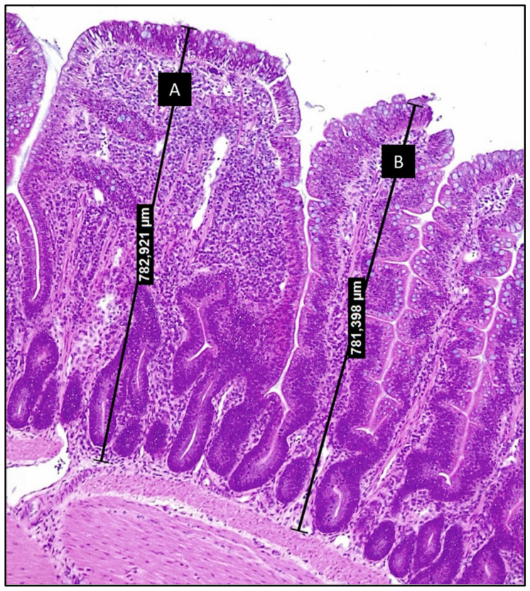
Figure 1.
Photomicrography of chicken ileum section stained with hematoxylin and eosin (200×) (from authors archive). Villi A and B display very close heights, although villus A presents increased lamina propria thickness and increased infiltration by inflammatory cells. These alterations are comprised by the ISI methodology and allow to relate structural alterations to the loss of functionality.

