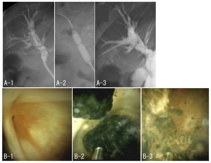
Figure 1.
Case 1. The beginning of the right anterior columnar canal was narrowed (B-1), and stones filled the upstream of the stenosis (A-1). Balloon dilation of the stenosis was performed (A-2). The stone was removed by electrohydraulic shockwave lithotripsy under peroral cholangioscopy (B-2,B-3), and cholangiography was performed to confirm the absence of residual stones (A-3).

