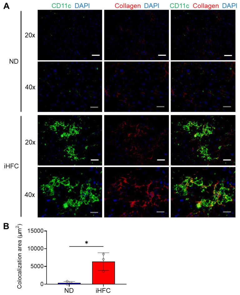
Figure 6.
The iHFC diet accumulates CD11c+ cells which are colocalized with collagen fibers. (A) Representative histological images (20× or 40× magnification) of fluorescent immunohistochemistry for CD11c, collagen type 1, and DAPI of the livers from TSNO mice on the ND or iHFC diet for 12 weeks. White scale bars, 100 μm. Gray scale bars, 50 µm. (B) Colocalization areas were calculated as described in Figure S10 and the Materials and Methods. Data are shown as means ± SD. * p < 0.05. Statistical significance was evaluated by unpaired Student’s t-test.

