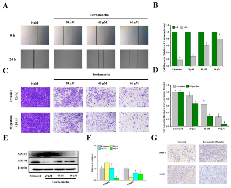
Figure 9.
Metastasis of Ishikawa cells and Ishikawa xenografts in mice treated with isorhamnetin. (A): Scratch healing of Ishikawa cells with different concentrations of isorhamnetin (0 μM, 20 μM, 40 μM, and 60 μM). (B): Quantification of the effects of isorhamnetin (0 μM, 20 μM, 40 μM, and 60 μM) on scratch healing. (C): Migration and invasion of Ishikawa cells treated with different concentrations of isorhamnetin (0 μM, 20 μM, 40 μM, and 60 μM). (D): Quantification of the migration and invasion of Ishikawa cells treated with different doses of isorhamnetin (0 μM, 20 μM, 40 μM, and 60 μM). (E): Effect of isorhamnetin on Ishikawa cell migration and invasion toward MMP2 and MMP9. (F): Quantification of the effects of isorhamnetin on MMP2 and MMP9 in terms of Ishikawa cell migration and invasion. (G): Immunohistochemistry of key proteins in Ishikawa xenograft mice treated with isorhamnetin (20 mg/kg). Different alphabetic letters (a, b, c, d) denote significant differences between different groups (p < 0.05, n ≥ 3).

