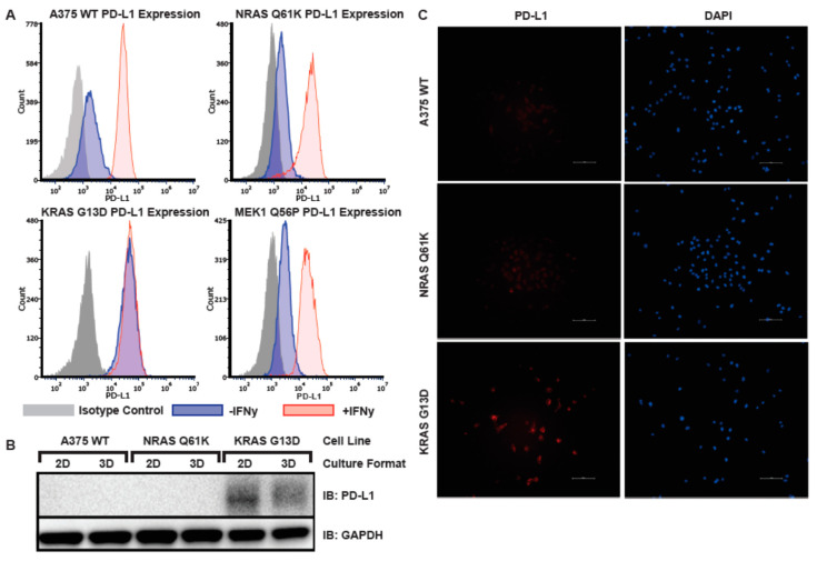
Figure 6.
PD-L1 is constitutively expressed in KRAS G13D, but not in A375 WT, NRAS Q61K, or MEK1 Q56P melanoma models. (A) Flow cytometry analysis of cell surface PD-L1 expression in A375 WT, NRAS Q61K, KRAS G13D, and MEK1 Q56P melanoma models grown in 2D tissue culture. Cells were treated overnight with 200 ng/µL interferon gamma (red), or mock treated (blue). The following day the cells were stained with either anti-PD-L1 or isotype control (grey); (B) PD-L1 immunoblot of total cellular protein from A375 WT, NRAS Q61K, and KRAS G13D cells grown in either 2D or 3D tissue culture; (C) Indirect immunofluorescence staining of PD-L1 in A375 WT, NRAS Q61K and KRAS G13D melanoma model lines.

