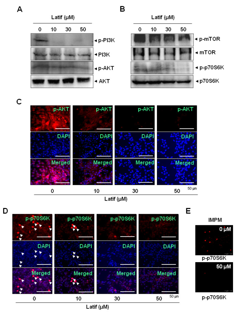
Figure 4.
Effects of Latif on the AKT pathway in human YD-10B OSCCs. (A,B) Amounts of 10, 30, and 50 μM Latif was treated for 24 h in YD-10B OSCCs, and then phospho-PI3K (p-PI3K), PI3K, phospho-AKT (p-AKT), AKT (A), phospho-mTOR (p-mTOR), mTOR, phospho-p70S6K (p-p70S6K), and p70S6K (B) were analyzed using Western blot analysis. β-actin level was detected as a loading control. (C–E) The cellular expression of p-AKT (C) and p-p70S6K (D,E) was analyzed using an immunofluorescence assay and images were observed using a fluorescence microscope (C,D) and validated using the intravital multi-photon microscope system (IMPM) for p-p70S6K (E). DAPI (blue) was used to stain the nucleus. Arrowheads: p-p70S6K- and DAPI-positive cells. Data are representative of the results of three experiments.

