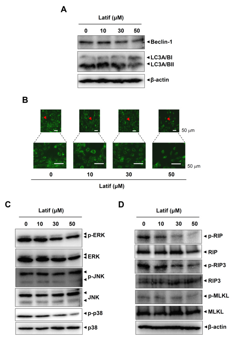
Figure 5.
Effects of Latif on autophagy and necroptosis in human YD-10B OSCCs. (A) Amounts of 10, 30, and 50 μM Latif was treated for 24 h in YD-10B OSCCs, and then Beclin1, LC3A/BI, and LC3A/BII expressions were assessed using western blot analysis. (B) Cells were treated with 10, 30, and 50 μM Latif, and then DAPGreen-positive autophagosome was observed using a fluorescence microscope. Arrowheads: Magnfied region. (C) Phospho-ERK1/2 (p-ERK1/2), ERK1/2, phospho-JNK (p-JNK), JNK, phospho-p38 (p-p38), and p38 levels were assessed using western blot analysis. (D) The cells were treated with Latif, and phospho-RIP (p-RIP), RIP, phospho-RIP3 (p-RIP3), RIP3, phospho-MLKL (p-MLKL), and MLKL were analyzed using Western blot analysis. The β-actin level was detected as a loading control. Data are representative of the results of three experiments.

