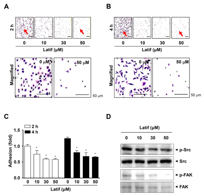
Figure 7.
Effects of Latif on cell adhesion and FAK/Src signaling in human YD-10B OSCCs. (A–C) Latif-treated cells (10, 30, and 50 μM) were seeded onto ECM-coated plates, and then the cell adhesion was monitored using a light microscope at the indicated time points, 2 h (A) and 4 h (B). Red arrows indicate the magnified region. The bar graph shows the relative value of cell adhesion (fold) normalized to the control (C). (D) Phospho-Src (p-Src), Src, phospho-FAK (p-FAK), and FAK were analyzed using Western blot analysis. Data are represented as mean ± S.E.M. Significance was set at p < 0.05, indicated by an asterisk in the graph.

