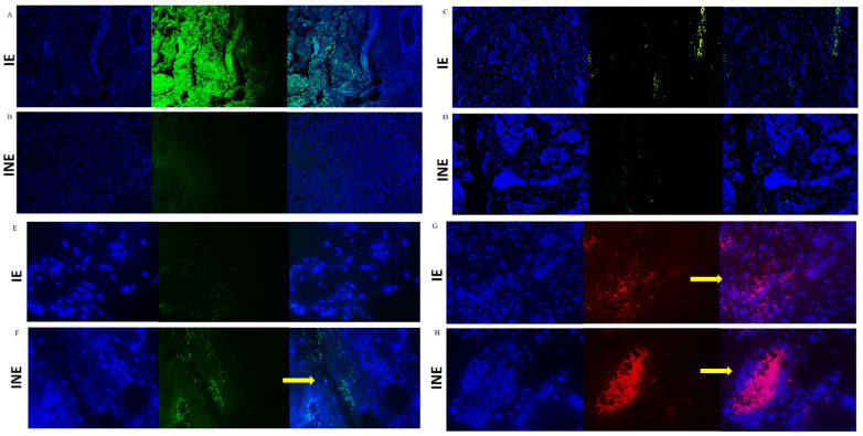
Figure 5.
Immunostaining of TGF-β1 (A,B), CTLA-4 (C,D), CD8+ lymphocytes (E,F), and CD4+ lymphocytes (G,H) in breast tumor biopsies from intermediate-risk patients exposed (IE) or unexposed (INE) to pesticides. Labeling was evaluated in breast tumors and infiltrating leukocytes (400×). The images sequentially represent (horizontal view) DAPI labeling, marker labeling, and the merge of DAPI + marker. The resulting images were merged in ImageJ to generate the final images. For all images: immunostaining in green for Alexa Fluor (positive staining), red for Texas Red (positive staining), and blue for DAPI (negative counterstaining). The yellow arrows indicate the labeled areas.

