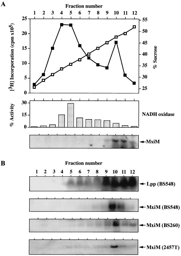
FIG. 4.
Subcellular localization of MxiM determined by sucrose density gradient centrifugation. Total cell envelope preparations were fractionated and analyzed as described in Materials and Methods. (A) Distribution of [3H]palmitate-labeled MxiM in samples derived from arabinose-induced BS548 cultures. Sucrose concentration (shown as open squares) and total 3H incorporation (shown as closed squares) are plotted against fraction number; NADH oxidase activity and labeled MxiM associated with each fraction are shown. (B) Immunological analysis of fraction components derived from unlabeled BS548 (grown with arabinose), BS260, and 2457T cultures. The positions of proteins recognized by either anti-Lpp or anti-MxiM serum are shown. Inner and outer membrane markers for BS260 and 2457T samples were indistinguishable from that described for BS548.

