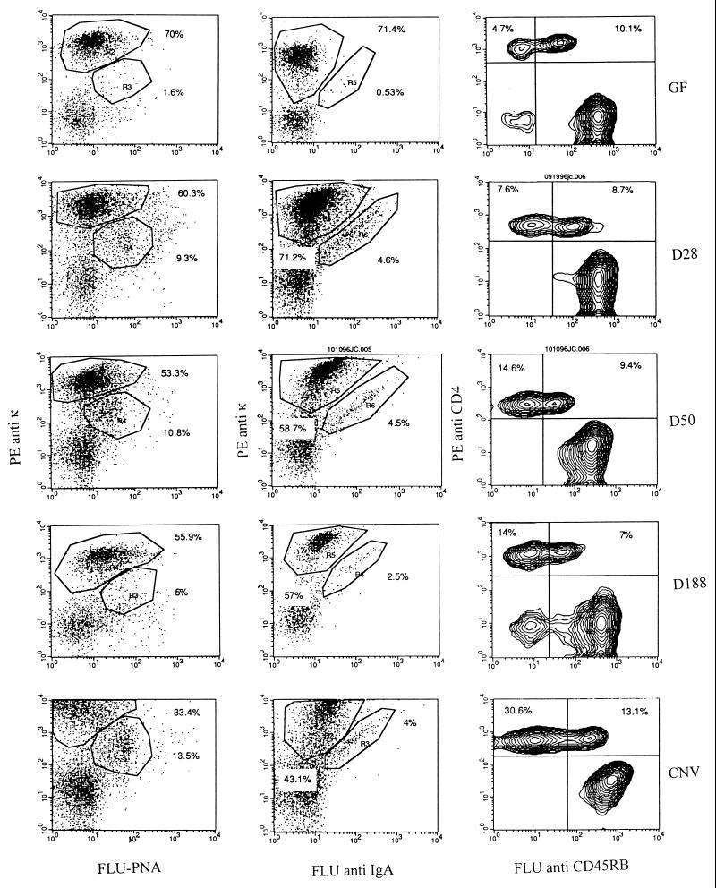
FIG. 1.
In order to assess the progress of germinal center reactions and activation of T cells in PP, cell suspensions from PP of three to five mice were analyzed by FACS analysis at various times following the colonization of formerly GF mice with SFB. These analyses were compared to those of PP from GF and conventionally reared (CNV) mice. Cells were stained with a germinal center marker, PNA, conjugated to FLU or FLU-labeled goat anti-mouse IgA, respectively, and then both were counterstained with PE-conjugated anti-kappa chain to monitor the development of germinal center reactions. Cells were also stained with the CD4 T-cell activation marker CD45RB, conjugated to FLU and counterstained with PE-conjugated anti-CD4 to monitor T-cell activation.

