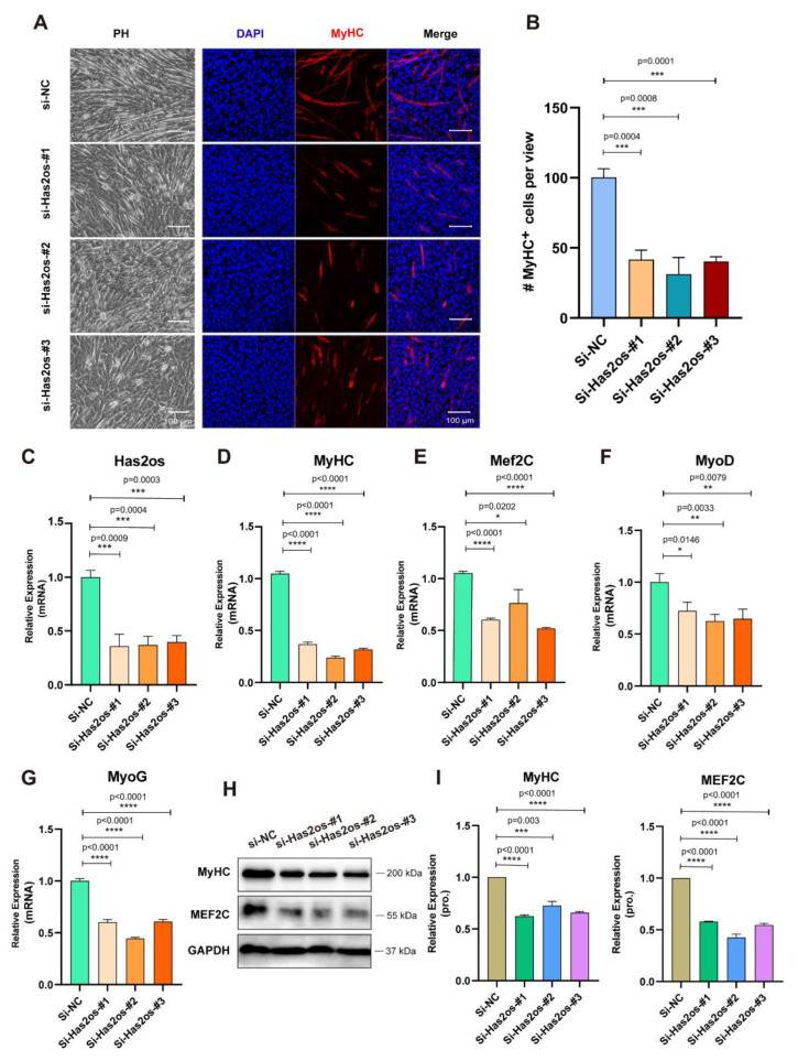
Figure 3.
Has2os knockdown inhibited myoblast differentiation. (A). Inhibition of myocyte fusion upon Has2os knockdown. Transfected cells were maintained in growth medium for two days and shifted into DM for differentiating another three days, then cells were photographed by microscope to show the morphological changes. Immunofluorescence microscopy analyzed the expression of myogenesis marker MyHC as red fluorescence in C2C12 cells transfected with si-Has2os or si-NC. Scale bar = 100μm. (B). MyHC+ cells were calculated based on staining described in panel (A). The results are normalized to those of with si-NC. (C). The qPCR results showed a successful Has2os knockdown by si-RNA. (D–G). The qPCR results showed the downregulation of the differentiation marker MyHC, Mef2C, MyoD, and MoyG after Has2os knockdown. (H). The Western blot results showed the downregulated myogenesis marker MyHC and MEF2C after Has2os knockdown. (I). Relative expression in (H) were calculated. GAPDH was the internal control. Values were presented as means ± SEM. The statistical significance was calculated by t-test.

