Figure 6.
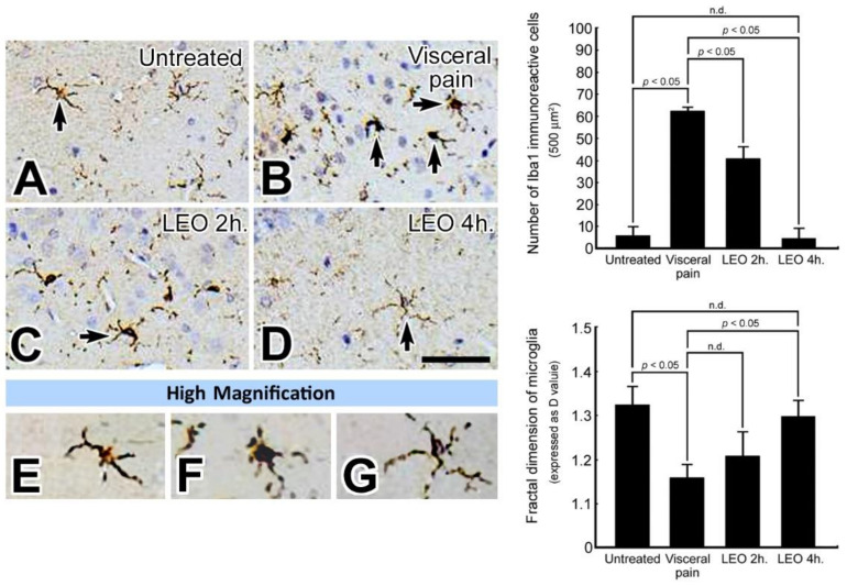
Photomicrographs (A–G) and histogram show the expression pattern of microglia (as determined by Iba1 immunohistochemistry) in the CeA of the untreated (A), visceral pain (B), and OS groups who received LEO for 2 h (C) and 4 h (D). Note that in the untreated group, only a small number of Iba1 immuno-reactive microglia with several branches and small cell bodies was detected in the CeA (arrow, A). However, following visceral pain, a large number of microglia with retracted branching and large cell bodies was detected in the CeA (arrows, B). Nevertheless, in rats subjected to visceral pain and who received LEO for 4 h, the expression pattern of microglia was much similar to that of untreated ones (arrow, D). High magnification (E–G) clearly demonstrates the functional status of the microglia in which a resting state was found in untreated (E) and OS group (G) while an activated state was detected in the group that suffered from visceral pain (F). Quantitative evaluation corresponded well with immunohistochemical findings in which OS successfully restored the branching pattern of microglia (as expressed by D value) following visceral pain. n.d. = no significant difference. Scale bar = 20 μm in (A–D), and = 10 μm in (E–G).

