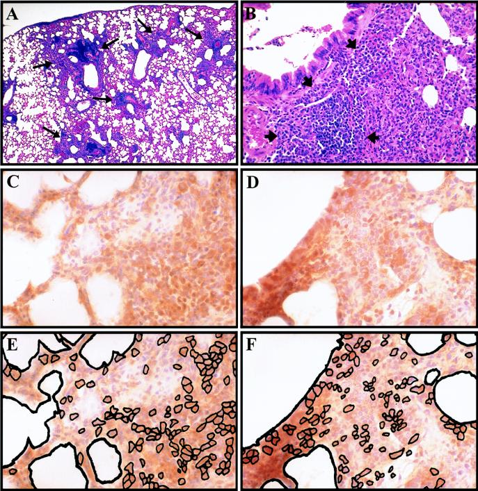
FIG. 1.
Lymphocytes in peribronchiolar lymphoid aggregates. Representative hematoxylin-and-eosin-stained sections (A and B) and sections immunohistochemically stained with hematoxylin counterstain (C to F) at 1 (A) and 3 (B to F) days postchallenge. (A) Lung of vaccinated mouse following challenge showing peribronchiolar lymphoid aggregates (long arrows); (B) higher magnification of peribronchiolar lymphoid aggregates (short arrows). Staining of adjacent sections for T lymphocytes (CD3+ cells) (C) and B lymphocytes (B220+ cells) (D) with corresponding schematic representation of stained cells within each section (E and F, respectively). Magnification: (A) ×25; (B) ×50; (C to F) ×100.

