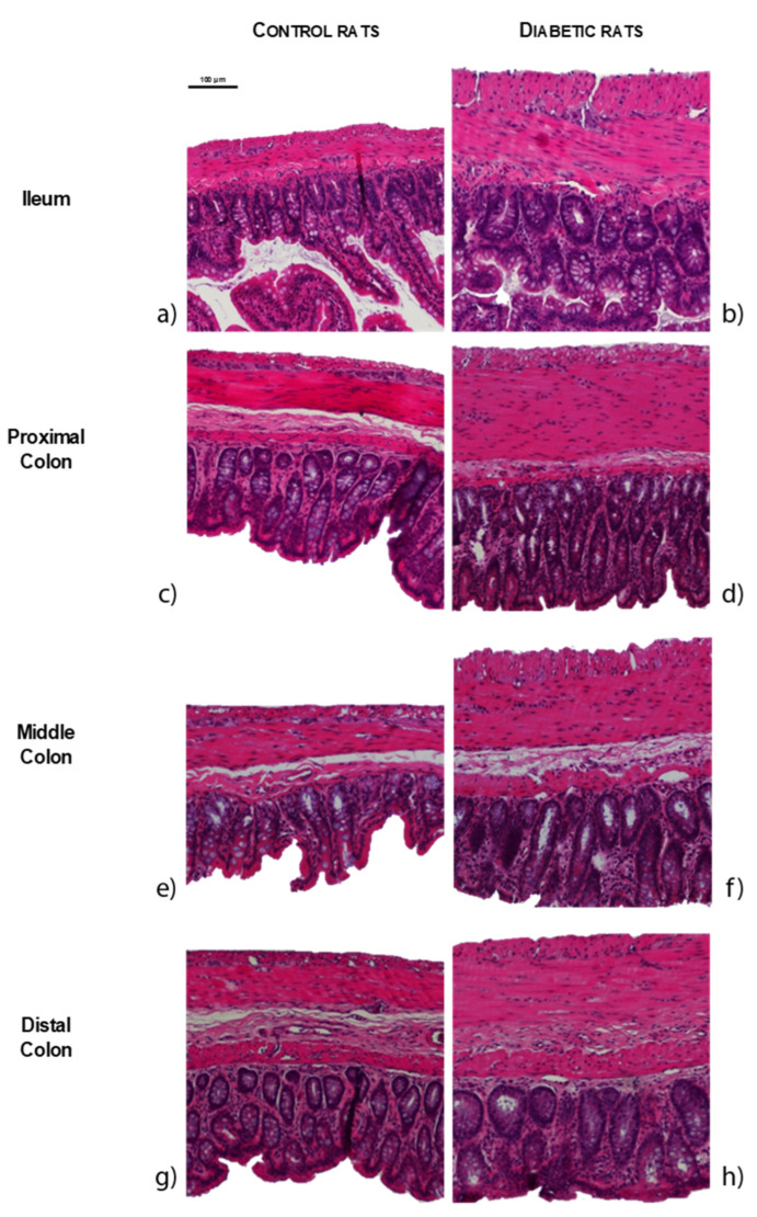
Figure 3.
Representative microscopic photographs of intestinal segments of control (CTRL, a,c,e,g) and streptozotocin-induced diabetic rats (STZ, b,d,f,h), stained with hematoxylin and eosin: ileum (a,b); proximal colon (c,d); middle colon (e,f) and distal colon (g,h). The scale bar (100 µm) is valid for all images.

