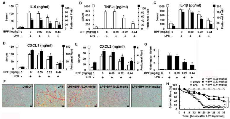
Figure 5.
BPF alleviates excessive cytokine production and tissue damage of the lungs and improves the survival rate in the LPS-induced mouse septic shock model. (A–E) Mice were injected with 2.5 mg/kg of LPS (intraperitoneal injection) and DMSO or BPF (0.09, 0.22 or 0.44 mg/kg, intravenous injection). After 4 h, the mice were euthanized. Blood and peritoneal fluid were collected and the levels of the indicated inflammatory cytokines were determined using ELISA (n = 5). (F,G) Mice were injected with 2.5 mg/kg LPS (intraperitoneal injection) and DMSO or BPF (0.09, 0.22 or 0.44 mg/kg, intravenous injection). After 24 h, the mice were euthanized and the left lung lobes were collected and fixed in formalin, followed by staining with H&E. H&E staining of lung tissues from each group was conducted, and representative images from three independent experiments conducted on three different days are shown. The bar represents 200 μm. Histopathological scores were obtained using an arbitrary scoring index based on the degree of inflammatory cell infiltration and the extent of the lesion area (n = 5). (H) Mice were injected with 25 mg/kg of LPS (intraperitoneal injection) and DMSO or BPF (0.09, 0.22 or 0.44 mg/kg, intravenous injection). The survival of the mice was monitored every 4 h for 36 h and the survival rates were expressed as a percentage (n = 20). All results are shown as the means ± SD of three different experiments with triple samples. * p < 0.5 compared to LPS only.

