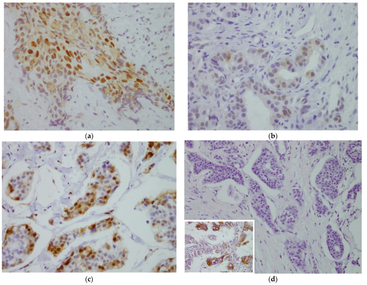
Figure 1.
Evaluation of immunohistochemical staining. (a) For HIF-1α, moderate staining of nuclei and slight staining of some cytoplasmic areas, >5% in tumor cells. 40×. (b) For pAKT, mild-to-moderate nuclear and cytoplasmic staining, ≥10% in tumor cells. 40×. (c) For pMAPK, strong nuclear staining and mild-to-moderate cytoplasmic staining, >10% in tumor cells. 40×. (d) For EGFR, negative membrane staining. A staining positive control for EGFR of lung cancer was inserted in the image 40×.

