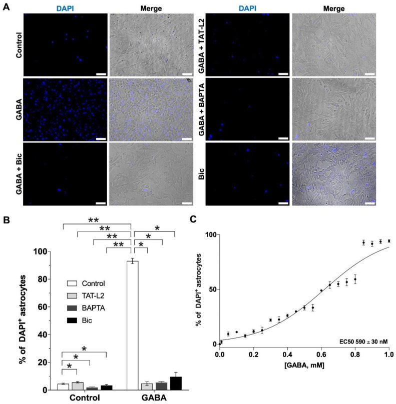
Figure 2.
GABA increases DAPI uptake in DI TNC1 astrocytes through Cx43 hemichannels via activation of GABAA receptors. (A) Representative photomicrographs showing DAPI uptake in DI TNC1 astrocytes under normal extracellular Ca2+ conditions (Control), show increased DAPI uptake in response to 1 µM GABA, effect that is prevented by preincubation with 100 µM bicuculline (Bic), 10 nM TAT-Cx43L2 (TAT-L2) or 100 nM BAPTA-AM (BAPTA). Scale bar: 50 µm. (B) Quantification of DAPI uptake. n = 3, with technical triplicates. (C) Concentration response curve for GABA on astroglial DAPI uptake performed to estimate the EC50 for GABA (EC50: 590 ± 30 nM). Error bars: SE; * p ˂ 0.05, ** p ˂ 0.01.

