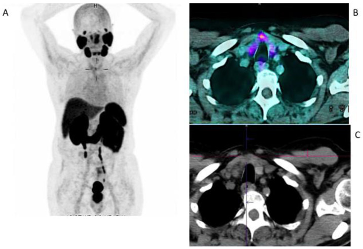
Figure 2.
Incidental findings of thyroid cancer in a 63-year-old patient studied with 68Ga-PSMA-11 for prostate cancer. MIP (A) and fused images (B) showing focal uptake (SUVmax 3.5) on the thyroid gland corresponding to a hypo-dense nodule on a co-registered low-dose CT scan (C). The ultrasound fine needle aspiration revealed Thy 4 (suspicious for malignancy) and the patient underwent a total thyroidectomy. The histology revealed a poorly differentiated papillary thyroid carcinoma.

