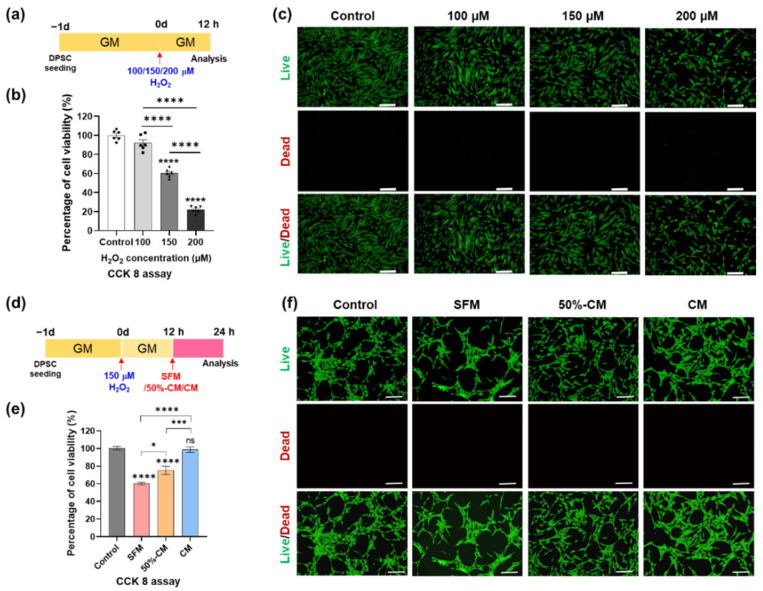
Figure 2.
Treatment with hGF-CM improves DPSC viability after H2O2 exposure. (a–c) DPSC viability exposed to different concentrations (100, 150, and 200 µM) of H2O2 for 24 h. (a) Schematic image of the timetable for the viability test depending on the H2O2 concentration. (b) Quantified results determined by CCK-8 analysis. (c) Representative live/dead images showing the oxidative stress effects induced by H2O2 treatment on DPSCs. (d–f) DPSC viability cultured in either CM, 50% CM, or SFM for another 12 h after exposure to 150 µM H2O2 for 12 h (d) Schematic image of the timetable. (e) Cell viability measured by CCK-8 analysis. (f) Representative live/dead images of DPSCs confirmed the antioxidative stress effect of hGF-CM. All data are represented as the mean ± SD. One-way analysis of variance followed (ANOVA) by Tukey’s multiple comparisons test was used to detect statistically significant differences between groups. Represents * p < 0. 05, *** p < 0. 001, **** p < 0.0001, ns = not significant, scale bar = 200 µm. SFM: Serum free medium, GM: Growth medium, CM: Conditioned medium, 50% CM: Conditioned medium diluted by 50% with serum free medium.

