Figure 9.
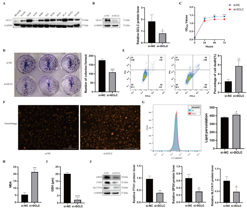
GCLC silencing promotes ferroptosis of H358 cells. (A) The expression level of GCLC in BEAS2B cells and NSCLC cell lines by Western blot. (B) After H358 cells were transfected with si-GCLC or si-NC for 72 h, the protein expression of GCLC was detected by Western blot assay. * si-GCLC vs. si-NC, * p < 0.05, ** p < 0.01, *** p < 0.001, ****p<0.0001. (C,D) CCK8 assay and colony formation assay were measured in siNC- or siGCLC-transfected cells. * si-GCLC vs. si-NC, * p < 0.05, ** p < 0.01, *** p < 0.001. (E) Cell death assay was determined by flow cytometry in H358 cells. * si-GCLC vs. si-NC, * p < 0.05, ** p < 0.01, *** p < 0.001. (F–I) Fe2+, lipid peroxidation, MDA, and GSH levels were examined in H358 cells transfected with si-NC or si-GCLC. * si-GCLC vs. si-NC, * p < 0.05, ** p < 0.01, *** p < 0.001. (J) The expression levels of GPX4, FTH1, and SLC7A11 in H358 cells with GCLC silencing were determined by Western blot. * si-GCLC vs. si-NC, * p < 0.05, ** p < 0.01, *** p < 0.001.

