Figure 8.
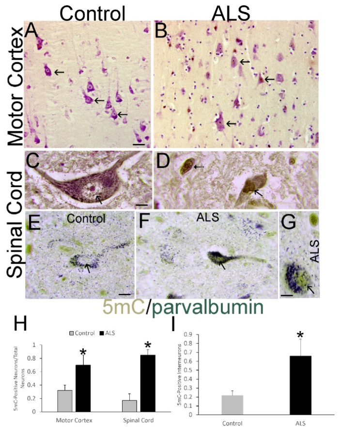
Upper MNs and spinal cord MNs and interneurons undergo hypermethylation in human ALS. (A) Layer V pyramidal neurons in control motor cortex (arrows) have low nuclear 5mC IR (brown). (B) In ALS motor cortex, numerous layer V pyramidal neurons (arrows) have nuclear 5mC IR (brown). (C) Spinal cord MNs in controls have chromatin threads (arrow) decorated with 5mC IR (brown) against a negative nucleoplasm. (D) Spinal cord MNs in ALS cases have shrunken nuclei that are strongly positive throughout for 5mC IR (arrow, brown) and cell bodies with somatodendritic attrition. These cells are discernible as attritional MNs because of the marginalized Nissl substance [62]. Other cells (hatched arrow) could be residual MNs or interneurons. (E) Interneurons, identified by parvalbumin (arrow, green/black) plus 5mC (brown), show scant DNA methylation IR in controls. (F,G) In ALS cases, parvalbumin (green/black, arrow) colocalizes with 5mC (brown). Interneuron in (G) is shown at higher magnification. Scale bars: (A) (same for (B)), 30 µm; (C) (same for (D)), 10 µm; (E) (same for (F)), 10 µm; (G), 5 µm. (H) Counts (mean ± SD) of motor cortical pyramidal neurons and spinal MNs with nuclear IR for 5mC in control (n = 6) and ALS (n = 8) cases. * p < 0.01, motor cortex; * p < 0.001, spinal cord. (I) Counts (mean ± SD) of parvalbumin-positive spinal cord interneurons with nuclear IR for 5mC in control (n = 6) and ALS (n = 8) cases. * p < 0.01.

