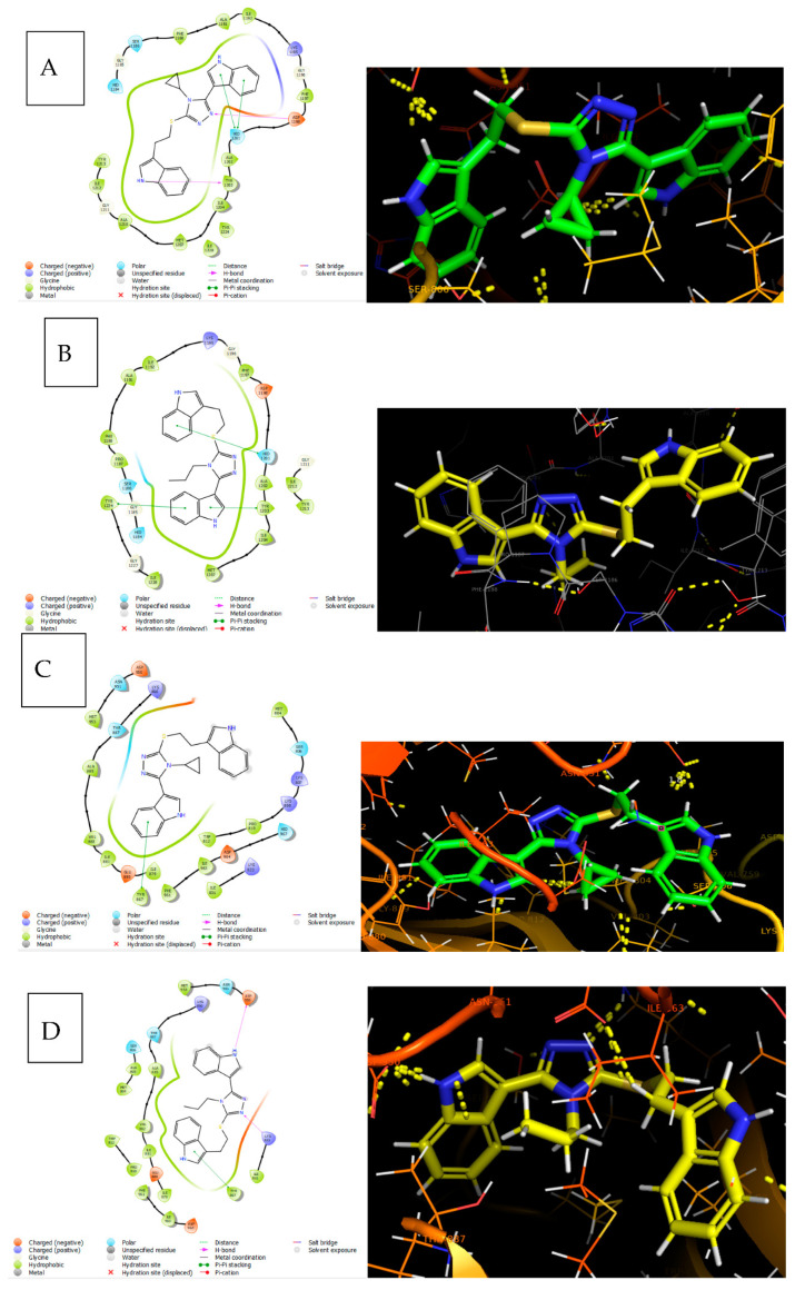
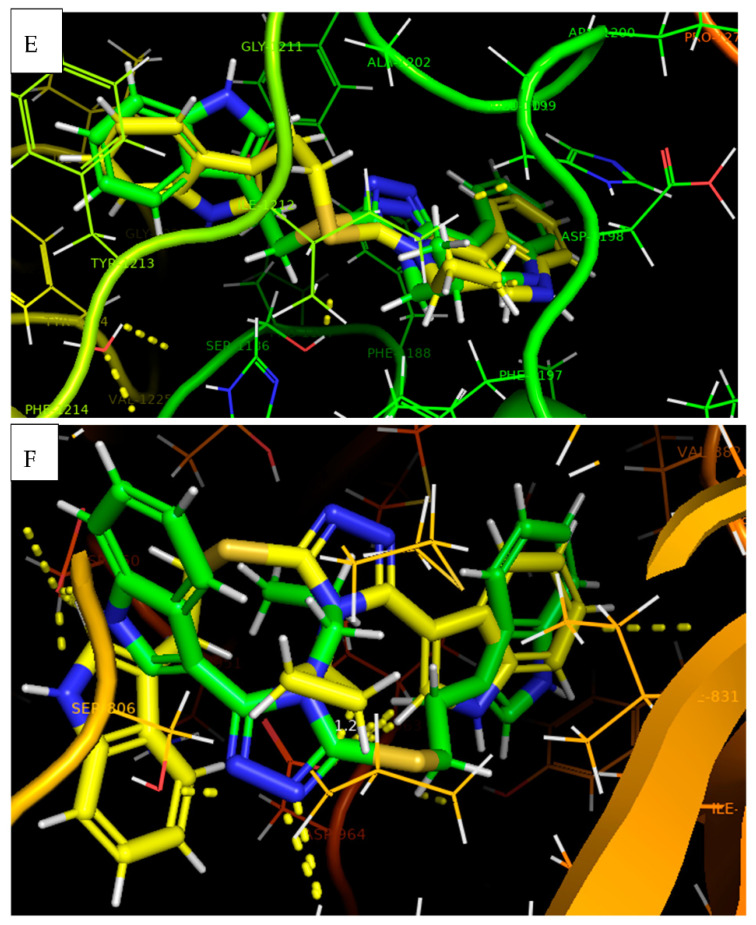
Figure 10.
(A) Docking pose of 15r in PDB ID 4OA7 shown in a 2D structure, where indole NH bonded with TYR 1203 via H-bonding, which is represented by the pink arrow line; similarly, triazole N bonded with ASP 1198. On the other hand, the indole shows a Pi–Pi stacking with HID 1201, represented by the green line. (B) Docking pose of 15o in PDB ID 4OA7 shown in a 2D structure, where both indoles show Pi–Pi stacking with HID 1201, TYR 1203, and TYR 1224, represented by the green line. (C) Docking pose of 15r in PDB ID 3L54 shown in a 2D structure, where indole shows Pi–Pi stacking with TYR 867, represented by the green line. (D) Docking pose of 15o in PDB ID 3L54 shown in a 2D structure, where indole NH and triazole N bonded with LYS 833 and ASP 950, respectively, via H-bonding, represented by the pink arrow line. On the other hand, the indole shows Pi–Pi stacking with TYR 867, represented by the green line. (E) Superimposable poses of 15r and 15o in PDB ID 4OA7 shown in a 3D structure. (F) Superimposable poses of 15r and 15o and standard in PDB ID 3L54 shown in a 3D structure. 15r and 15o are represented by green and yellow colours.


