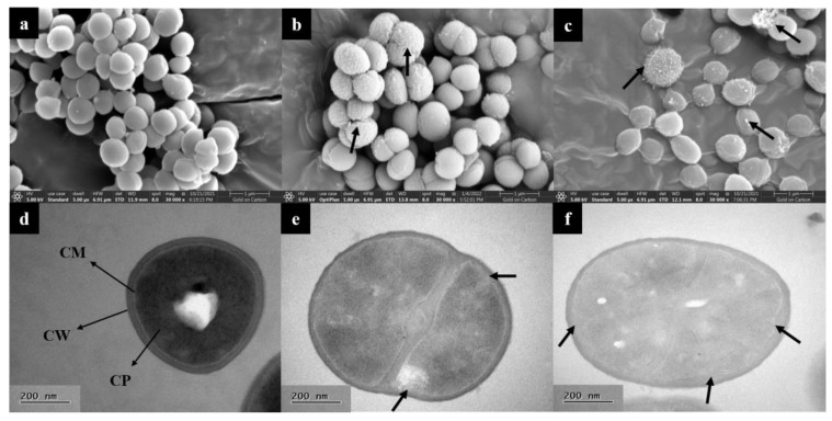
Figure 4.
Electron microscopy analysis of S. aureus. SEM images of S. aureus treated with L-36 CFCS ((a) CK; (b,c) 1 × MIC). Stubby black arrows: intracellular substances and damaged cell. TEM images of S. aureus treated with L-36 CFCS ((d) CK; (e,f) 1 × MIC). CW: cell wall; CM: cell membrane; CP: cytoplasm. Stubby black arrows: membrane invagination and damaged.

