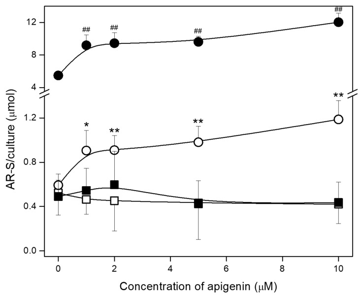
Figure 4.
The effect of apigenin on the mineralization of hFOB 1.19 and Saos-2 cells. Quantitative analysis of calcium deposits in hFOB 1.19 (squares) and Saos-2 (circles) cells under resting conditions (open squares/circles) or in the presence of stimulators, AA, and β-GP (filled squares/circles) was carried out by staining with AR-S, de-staining with CPC, and absorbance measurements at λ 562 nm. The degree of mineralization was normalized to the relative number of viable cells. Data are means ± S.E. of at least three independent experiments (* p < 0.05, **/## p < 0.01 compared with resting/stimulated cells without apigenin).

