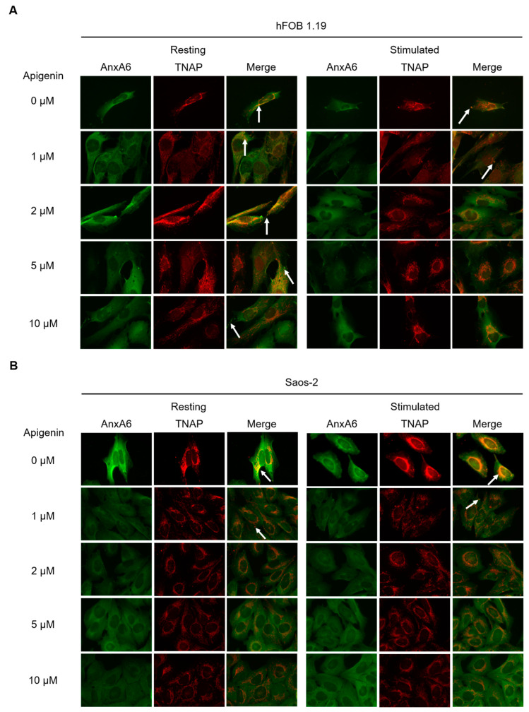
Figure 6.
Co-localization of AnxA6 and TNAP during mineralization of hFOB 1.19 (A) and Saos-2 (B) cells under resting conditions or in the presence of stimulators, AA and β-GP. Cells were incubated with different concentrations (µM) of apigenin for 7 days, fixed and analyzed by fluorescent microscopy (magnification 630×). AnxA6 (green) was immunostained with anti-AnxA6 primary antibody conjugated with Alexa Fluor 488 secondary antibody. TNAP (red) was immunostained with anti-TNAP primary antibody conjugated with Alexa Fluor 594 secondary antibody. Sites of AnxA6 and TNAP co-localization are visible in yellow on merge images (arrows).

