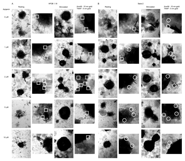
Figure 8.
TEM images of the co-localization of AnxA6 and TNAP in MVs during the mineralization of hFOB 1.19 (A) and Saos-2 (B) cells under resting conditions or in the presence of stimulators, AA and β-GP. Cells were incubated with different concentrations (µM) of apigenin for 7 days, lysed, and analyzed by TEM (magnification 100,000×). Bar: 200 nm. Additional magnifications 300,000×. AnxA6 was labelled with anti-AnxA6 primary antibody conjugated with 10 nm colloidal gold secondary antibody. TNAP was labelled with anti-TNAP primary antibody conjugated with 5 nm colloidal gold secondary antibody. Sites of AnxA6 and TNAP co-localization are marked by squares for hFOB 1.19 cells and circles for Saos-2 cells.

