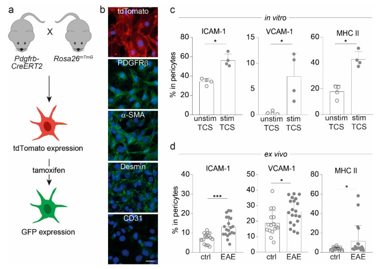
Figure 1.
Pericytes from Pdgfrb-CreERT2-GFP mice express molecules important for cell adhesion and antigen presentation. (a) Overview of Pdgfrb-CreERT2-GFP mice with tdTomato-labeled pericytes, which express GFP upon tamoxifen administration. (b) Images of cortical pericytes stained with immunofluorescent antibodies depict the expression of tdTomato, typical mural cell markers (PDGFRβ, α-SMA and desmin), and CD31 as endothelial cell marker (scale bars = 50 µm). (c) Bar graphs show the frequencies of ICAM-1, VCAM-1 and MHC II positive cells in pericytes treated with T cell supernatant (TCS) either from 72 h-stimulated or unstimulated T cells in vitro after 72 h assessed via flow cytometry. (d) Frequencies of ICAM-1, VCAM-1, and MHC II-expressing pericytes isolated from the CNS of Pdgfrb-CreERT2-GFP animals immunized with MOG35–55 at disease maximum (EAE; n = 20) in comparison to non-immunized mice (ctrl; n = 16) are illustrated. All data depict mean ±SD. Whereas one dot represents for (c) one individual experiment and for (d) one dot represents one individual mouse. Data shown are from at least three independent experiments. Statistical analysis was performed using Mann–Whitney U-test. *, p ≤ 0.05; ***, p ≤ 0.001.

