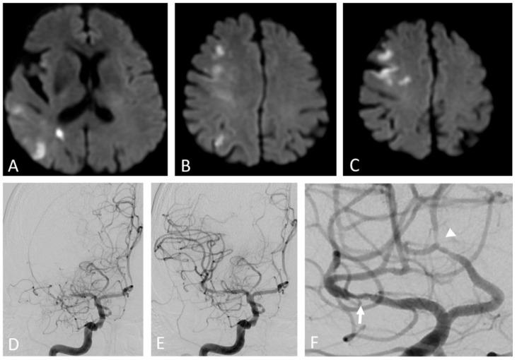
Figure 3.
Artery-to-artery embolism and in situ thrombotic occlusion of the middle cerebral artery due to intracranial atherosclerotic disease. Hypertensive 67-year-old man. Diffusion-weighted magnetic resonance imaging demonstrates multiple cortical-subcortical ischemic lesions in the territory of the right middle cerebral artery (A–C). Digital subtraction angiography shows an occlusion of the M1 segment of the right middle cerebral artery (D) and its complete recanalization after mechanical thrombectomy (E). The magnified oblique projection after the recanalization (F) reveals an underlying atherosclerotic plaque at the site of the previous occlusion (arrow) and additional stenotic lesions along the course of the right anterior cerebral artery (arrowhead).

