Figure 2.
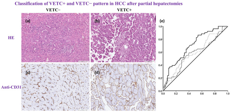
Histological photo based on Hematoxylin & Eosin stain or Anti-CD31; ROC curves for predicting VETC+. All surgical specimens were sent to and processed by a qualified pathologist. (a,b), H&E staining is the gold standard for medical diagnosis. Hypercellular density was noted in the VETC+ group, but the vascular structure was not clear on the H&E stain. (c,d), special anti-CD31 stain for the evaluation of intra-tumoral endothelial cells. The number of CD31-positive vessels/mm2 indicated intra-tumoral MVD. There were two distinct vascular patterns: (c), capillary-like vessels and disconnected blood vessels with small or no lumen indicated VETC−; (d) vessels that encapsulated tumor clusters and formed a cobweb-like pattern indicated VETC+; scale bar, 100 μm. (e) ROC curves for predicting VETC+ according to intra-tumoral MVD, the MVD of the normal part of the liver, and serum α-fetoprotein (black line, dotted line and grey line, respectively). Only intra-tumoral MVD independently predicted VETC+ (AUROC = 0.693; 95% CI, 0.613 to 0.780, p < 0.001). When the cut-off score was 40 vessels/mm2, the sensitivity and specificity of VETC+ was 0.863 and 0.461, respectively. Anti-CD31, cluster of differentiation 31 antibody; ROC curve, receiver operating characteristic curve; VETC+, positive VETC pattern; H&E stain, Hematoxylin & Eosin stain; MVD. Microvessel density; VETC−, negative VETC pattern, AFP, α-fetoprotein; Intra-tumoral MVD, black line; Normal part liver MVD, dotted line; Serum AFP, grey line.

