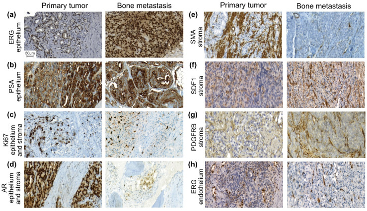
Figure 1.
Sections from paired primary prostate tumors and bone metastases immune stained for ETS-related gene (ERG) (a,h), prostate specific antigen (PSA) (b), the marker for proliferation Ki67 (c), androgen receptor (AR) (d), smooth muscle actin (SMA) (e), stroma derived factor 1 (SDF1) (f), and platelet-derived growth factor receptor β (PDGFRB) (g), all seen at the same magnification (see scalebar in (a)). (a) ERG positive tumor cells in a primary tumor and in the paired metastasis. (b) Moderate (left side) to high (right side with glands) PSA staining in a primary tumor. Moderate PSA staining and glandular differentiation was seen in the metastasis (classified as MetA). (c) Sections with relatively high Ki67 labeling in epithelium and stroma in a primary tumor and in the paired bone metastasis. (d) AR positive epithelial and stroma cells were seen in the primary tumor. In the metastasis (classified as MetA), a nest of AR positive tumor cells was growing in a bone marrow space and a few AR positive cells are seen on the bone surface. (e) Numerous SMA positive smooth muscle cells are seen in the primary tumor, whereas only few cells were SMA positive in the paired bone metastasis (here only in blood vessel walls). This metastasis, lacking bone structures, was classified as MetB. (f) SDF-1 positive cells were seen in the stroma in the primary tumor, mainly in blood vessel walls. Such cells were also seen in the bone metastasis stroma. (g) PDGFRB positive stroma cells, mainly in blood vessel walls and in fibroblasts, were seen in the primary tumor stroma and in the paired metastasis (classified as MetB). (h) ERG positive endothelial cell nuclei were seen in the primary tumor and in its bone metastasis. Note that several tumor cells are lying close to endothelial cells. In this case, the tumor cells were ERG negative. This metastasis was classified as Ki67 high/PSA low but with unknown MetA-C status. Blood vessels with ERG positive endothelial cells and walls positive for SMA, PDGFR-beta, and SDF-1 were a dominant component of the bone metastasis stroma.

