Figure 1.
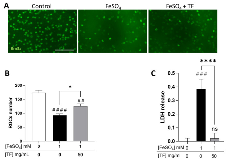
Transferrin protects RGCs in iron-treated retinal explants. (A) Representative images of retinal whole-mounts prepared for Brn3a immunostaining following 24 h incubation with 1 mM FeSO4 or transferrin (TF) (50 mg/mL) combined with FeSO4 and further cultured for 72 h (96 h total) in presence or not of TF. Untreated explant served as control. (B) Brn3a-positive cells were counted, showing that TF significantly protects against the loss of RGCs from iron overload. (C) LDH released by explants during 3 h to 24 h of culture was significantly increased in presence of 1 mM FeSO4 but absent in additive presence of TF. Data represent means ± SEM, n = 8–9 explants per condition. Statistical analysis was performed with Kruskal–Wallis and Dunn’s tests for multiple comparisons. ns: not significant; ## p < 0.01; ### p < 0.001; #### p < 0.0001 compared to control; * p < 0.05; **** p < 0.0001; compared to stress condition. Scale bar: 100 µm.

