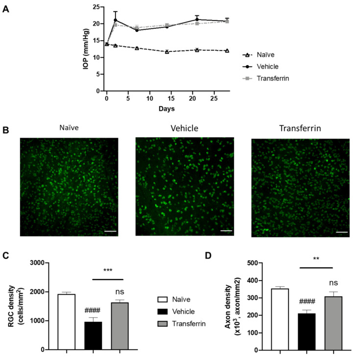
Figure 4.
Transferrin prevents retinal ganglion cell death and axon loss in rats exposed to ocular hypertension. (A) Rats were injected with microbeads in the anterior chamber on day 0. IOP remained significantly elevated over the four-week period of the study beginning at 48 h after OHT when compared to normotensive naïve animals. IOPs were not statistically different in vehicle- and TF-treated groups. (B) Representative micrographs of flat-mounted retina sections labeled for the RGC marker RBPMS. (C) Mean RGC density per mm2 in the vehicle group was significantly reduced when compared with control animals (naïve group). TF significantly preserved RGC. (D) Axon density in optic nerves from vehicle-treated group was significantly lower than naïve. There was no difference in axon density between the TF treatment and naïve optic nerves. Data represent means ± SEM, n = 10 per group. Statistics are one-way ANOVA followed by Tukey’s multiple comparisons test. Symbols above the bars are comparisons with naïve control group. ns, not significant; #### p < 0.0001 compared to naïve control group. ** p < 0.01; *** p < 0.001 compared to vehicle-treated group.

