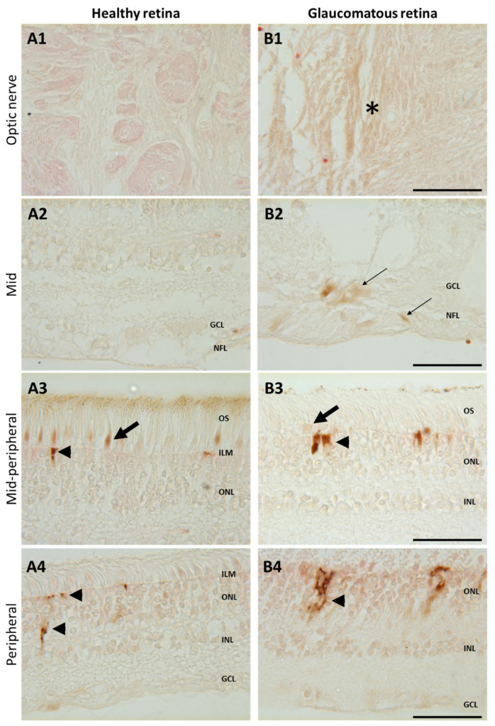
Figure 5.
Increased iron in glaucomatous versus healthy human retinas. Representative images of enhanced Perls’ reaction realized on human healthy eyes (A) and glaucoma-affected eyes (B) observed in the optic nerve (A1, B1), in the mid- and mid-peripheral retina (A2–3,B2–3), and in the peripheral retina (A4,B4). In healthy retina, iron was localized in cone outer segments ((A3), arrows) and in the Müller glial cell end-feed at the inner limiting membrane (ILM) ((A3), arrowhead) and in processes ((A4), arrowhead). Glaucomatous retinas present iron deposits in optic nerve ((B1), asterisk) and in the nerve fiber and ganglion cell layers ((B2), thin arrow) and increased iron staining in Müller cells ((B3,B4), arrowheads) but decreased in cone outer segments ((B3), arrow). Scale bar: 50 µm. GCL: Ganglion cell layer; ILM: Inner limiting membrane; INL: Inner nuclear layer; NFL: Nerve fiber layer; ONL: Outer nuclear layer. OS: Outer segment.

