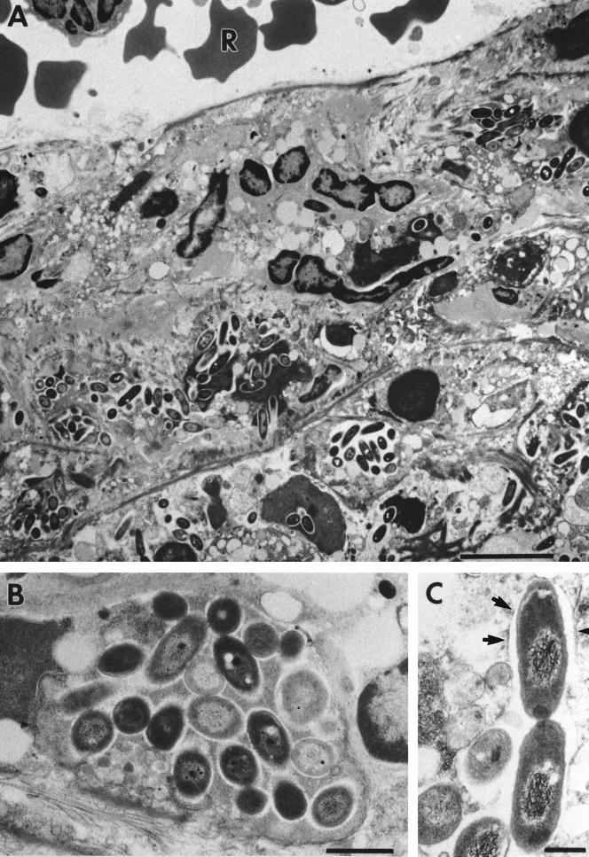
FIG. 5.
Electron microscopic investigation of spleens of C57BL/6 mice at day 6 after infection with 2 × 10² CFU of mouse-passaged strain B. pseudomallei NCTC 7431. Ultrathin sections through lesions of the spleen are shown. Within these lesions, we consistently detected the accumulation of B. pseudomallei (A). (B) Higher magnification of panel A showing densely packed bacteria. (C) Numerous intracellularly dividing bacteria surrounded by phagosomal membranes (arrows). R, erythrocyte; Bars represent 5 μm in panel A, 1 μm in panel B and 0.5 μm in panel C.

