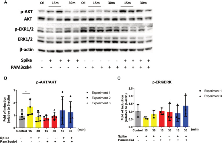
Figure 3.
SARS-CoV-2 S protein triggered the induction of the PI3K/Akt pathway. A549 cells were treated with 50ng/ml SARS-CoV-2 S protein or PAM3csk4 or SARS-CoV-2 S and PAM3csk4 for 15 and 30 minutes. SARS-CoV-2 S protein slightly decreased the phosphorylation of ERK1/2 (A, C), while significantly increased the phosphorylation of AKT (A, B). Representative western blot (A) is presented and densitometry analysis from three (B) or two (C) independent experiments. Densitometry analysis is illustrated in bar graphs and plotted as mean ± S.D. Individual points indicate the biological replicates from all experiments (experiments 1 and 2 include two biological replicates for each condition and experiment 3 includes one biological replicate per condition). Statistical analysis was performed with t test (B), but it was omitted for ERK due to the small sample size (C). *p < 0.05.

