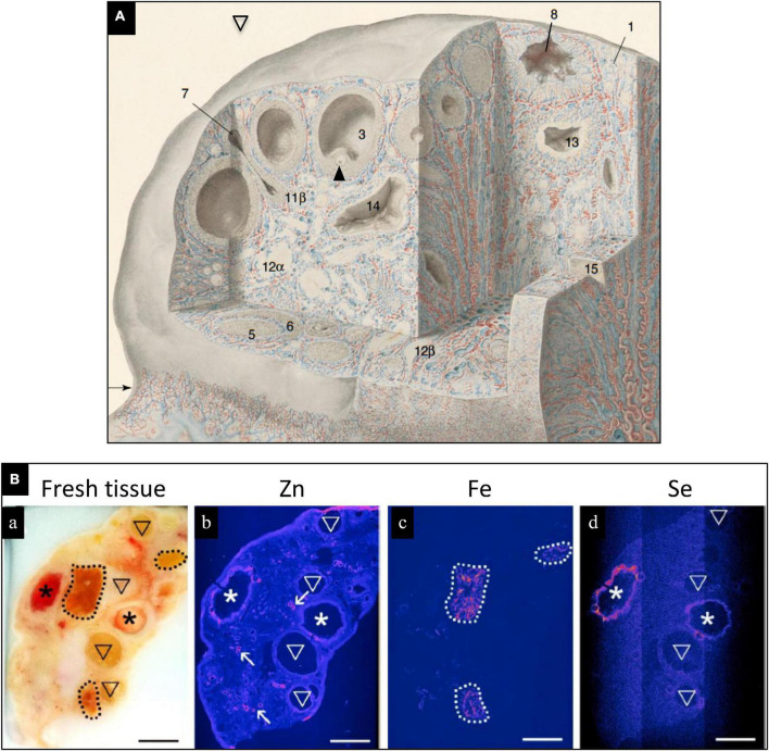
FIGURE 1.
(A) Drawing of human ovary with sections removed to reveal histological details of an antral (preovulatory) follicle (3) containing an oocyte (arrowhead) and a postovulatory follicle that has released its oocyte and partially collapsed (8). Blood vessels are colored red (arteries) and blue (veins); modified, with permission, from Clark, 1900. (B) Trace elements localized in bovine ovaries by synchrotron x-ray fluorescence (S-XRF). (a) Represents fresh tissue. Zinc (b, pink) localized primarily to blood vessels, Fe (c, pink) localized primarily to corpora lutea, and Se (d, pink) localized to healthy, preovulatory follicles (*) but not to atretic (regressing) antral follicles. Modified, with permission, from Ceko et al. (6).

