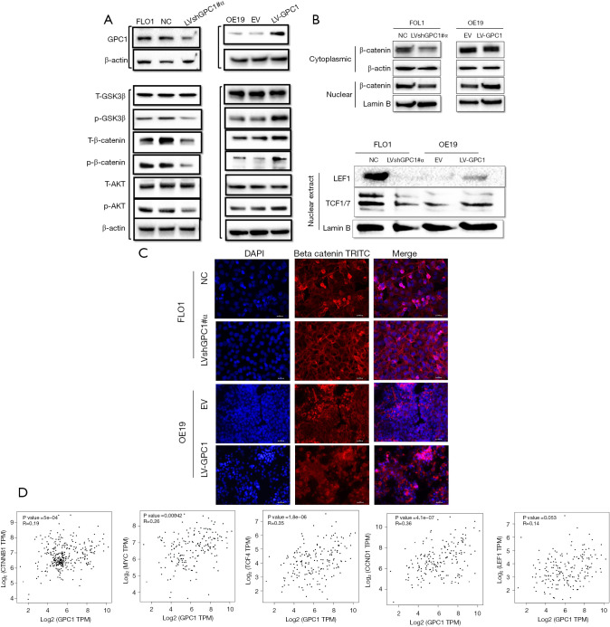
Figure 10.
GPC1 activates AKT/GSK-3β/β‐catenin pathway. (A) Western blot analysis of key proteins of AKT/GSK-3β/β‐catenin pathway. (B) Western blot assay examining the nuclear and cytoplasmic expression of catenin (left) and nuclear expression of β‐catenin downstream target transcription factors (LEF and TC-F1) in GPC1 knockdown FLO1 and GPC1 overexpressed OE19 cells. (C) Dual immunofluorescence staining for β‐catenin (red) and nucleus with DAPI (blue) was performed in GPC1 knockdown FLO1 and GPC1 overexpressed OE19 cells. Compared to controls, knockdown of GPC1 showed increased membrane staining and reduced nuclear staining (pink) of β‐catenin, while GPC1 overexpression resulted in reduced membrane staining and increased nuclear accumulation (pink) in OE19 cells. Scale bar: 50 µm. (D) The expression of GPC1 correlated with β‐catenin, c-myc, TCF4, cyclin D1, and LEF1 levels which were analyzed in the GEPIA database. Lamin B and β-actin were used for loading controls nuclear and cytoplasmic fractions respectively. NC, negative scrambled control; LVshGPC1#α, GPC1 knockdown plasmid; EV, empty vector control; LV-GPC1, overexpressing GPC1 lentivirus; DAPI, 4',6-diamidino-2-phenylindole-stained DNA; TRITC, tetramethylrhodamine; TPM, transcripts per million; GPC1, glypican 1.

