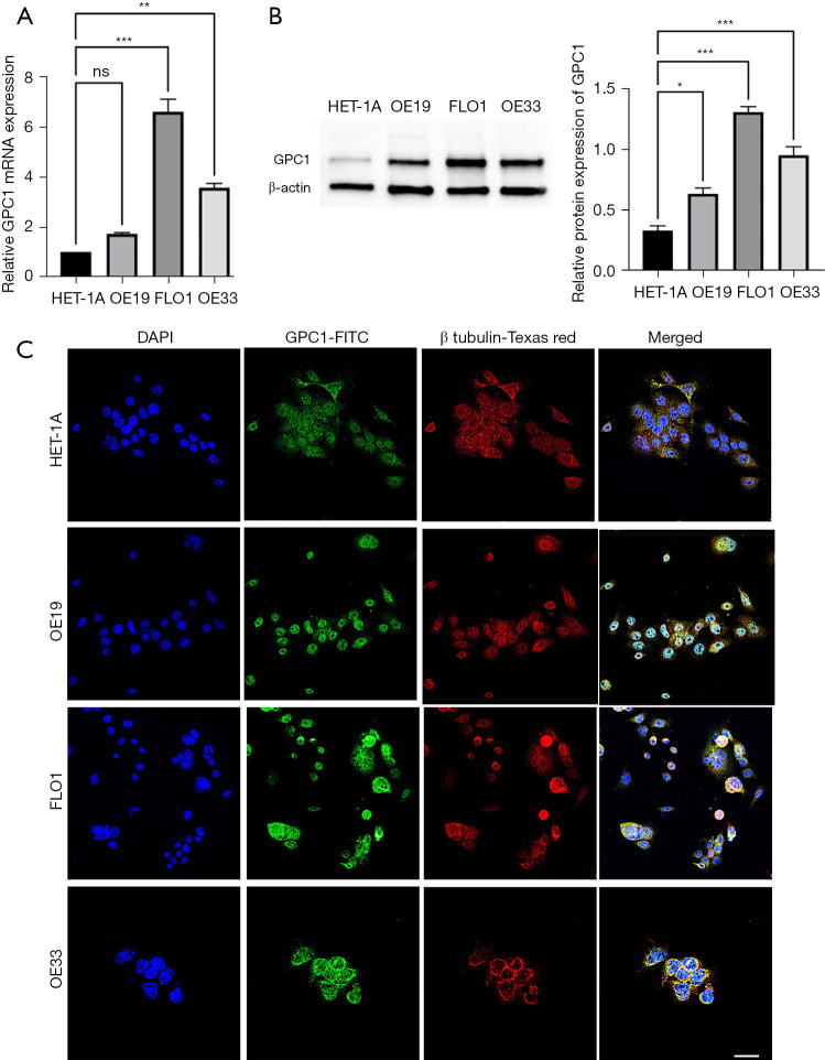
Figure 2.
GPC1 is highly expressed in esophagogastric cell lines. (A) Real time qPCR mRNA expression of GPC1 in HET-1A and EGAC cell lines (OE19, FLO1 and OE33). (B) Western blot and densitometry analysis of GPC1 protein in HET-1A and EGAC cell lines (OE19, FLO1 and OE33). Data represent mean ± SD. n=3. *, P<0.05, **, P<0.005, ***, P<0.001 vs. HET-1A. (C) Confocal images showing GPC-1 protein (green) predominantly in cytoplasm. Co-localization of GPC1 (orange-yellow staining) with cytoplasmic organelles. No expression was detected in nucleus or nuclear membrane. Green marks GPC-1, red marks β tubulin and blue marks DAPI-stained DNA. Scale bar: 100 µm. GPC1, glypican 1; ns, not significant; DAPI, 4',6-diamidino-2-phenylindole; FITC, fluorescein isothiocyanate; qPCR, quantitative polymerase chain reaction; EGAC, esophagogastric cancer; HET-1A, normal human epithelial cell line; SD, standard deviations.

