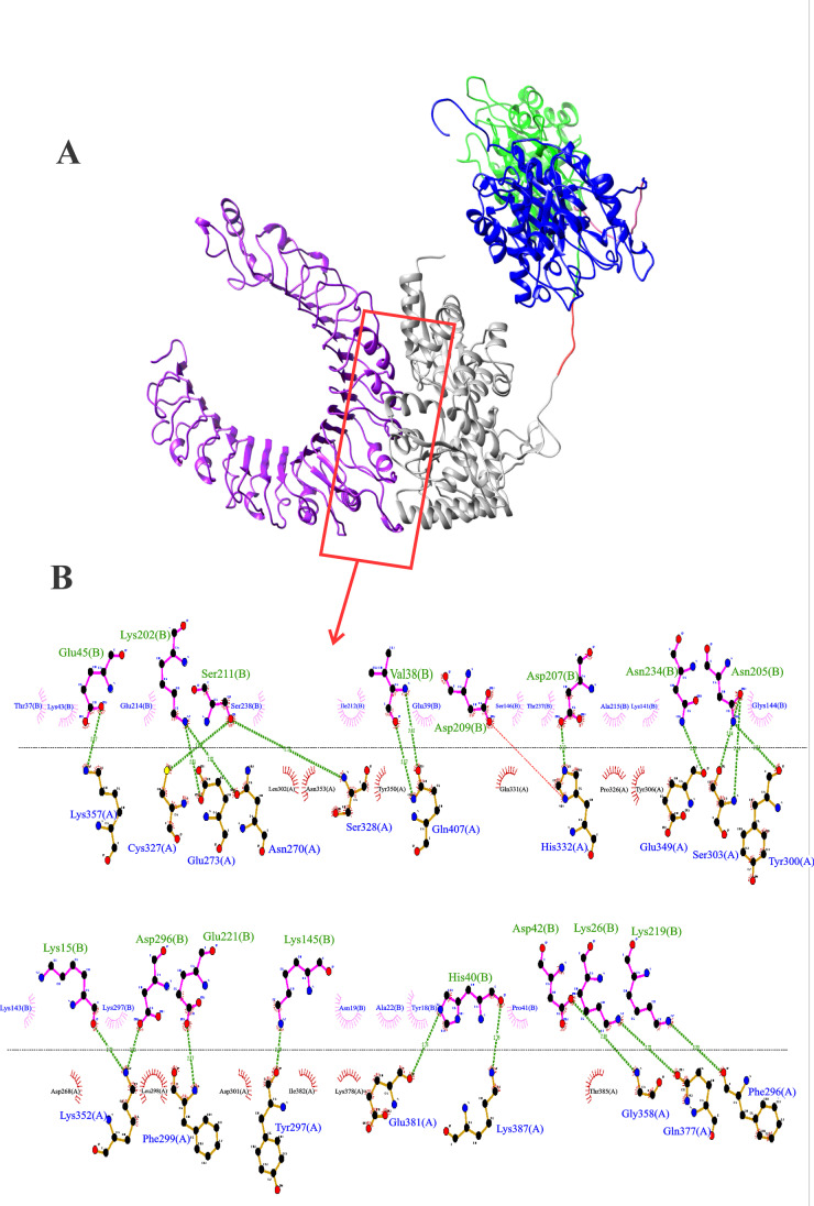
Fig. 2.
Anchoring of the MBP:PLD:CP40 fusion protein to the TLR2 receptor. A Structure generated by the Chimera software; TLR2 (receptor) in purple and MBP:PLD:CP40 protein (ligand) with MBP (grey), TEV protease site (red), PLD (green) rigid linker (pink), and CP40 (blue). B Residue interactions between proteins provided by the LIGPLOT v.2.2 software. Green lines represent hydrogen bonds, semicircles denote hydrophobic interactions. It can be seen 21 hydrogen bonds and 29 hydrophobic interactions between MBP and the TLR2. The MBP aminoacid envolved in the hydrogen bonds are written in green and followed by the letter (B), and the TLR2 residues envolved in these bonds are written in blue and followed by the letter (A). The MBP and TLR2 amino acid residues that participate in the hydrophobic interactions are represented inside pink and red semicircles, respectively

