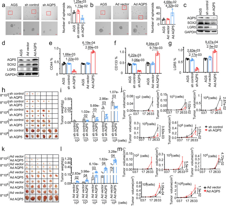
Fig. 3.
AQP5 promotes the malignant biological function of GC-CSCs in vitro and in vivo. a and b Representative images of exogenous AQP5 knockdown (a) or AQP5 overexpressing (b) AGS cells cultured in serum-free medium for 10 days. Statistical analysis was performed on the number of spheroids (diameter > 50 μm). (c and d) The expression levels of AQP5, LGR5 and SOX2 were measured using WB analyses after AQP5 overexpression and knockdown. e–g Flow cytometric analysis of CD44, CD133 and LGR5 expression in AQP5-overexpressing or AQP5-knockdown HGC-27 cells. h-m AQP5 was knocked down (h) or overexpressed (k) in HGC-27 cells. These cells were diluted and subcutaneously injected into severely immunodeficient mice. Tumors were examined over a 33-day period (n = 6 for each group). The tumor weight (i, l) and tumor volume (j, m) were monitored in the indicated groups and at the indicated time points

