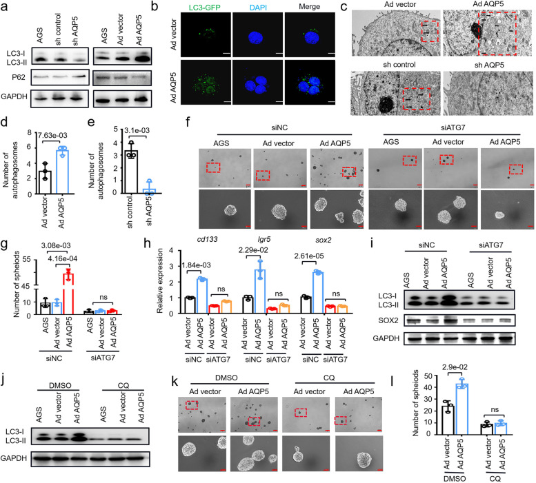
Fig. 5.
AQP5 activates GC-CSCs by inducing autophagy. a The expression levels of LC3 and p62 were evaluated in AQP5-knockdown or AQP5-overexpressing AGS cells. b AGS cells was infected with an adenovirus that expressed GFP-linked LC3 (GFP-LC3). Confocal microscopy was used to obtain fluorescent images. c TEM was used to assess autophagosome formation in AQP5-knockdown or AQP5-overexpressing AGS cells (red dashed boxes, black arrows indicate autophagolysosomal structures). d and e Statistical analysis of observed autophagosomes. f The spheroid-forming ability was evaluated after transfection of ATG7 siRNA in AQP5-overexpressing AGS cells. g Statistical analysis was performed on the number of spheroids (diameter > 50 μm). h The expression levels of cd133, lgr5 and sox2 in AQP5-overexpressing AGS cells were assessed after transfection with ATG7 siRNA. i Expression of LC3 and SOX2 in AQP5-overexpressing AGS cells was assessed after transfection of ATG7 siRNA. j LC3 expression in AGS cells overexpressing AQP5 was analyzed after CQ treatment. k The spheroid-forming of AGS cells overexpressing AQP5 was assessed after CQ treatment. l Statistical analysis was performed on the number of spheroids (diameter > 50 μm)

