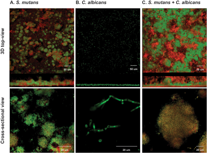
Figure 2.

Morphogenesis of micro-colonies in 48-h biofilms (1% sucrose condition) In 1% sucrose condition, the 48-h biofilms were visualized by a two-photon laser confocal microscope. The biofilms of single species were shown in panel A (S. mutans) and panel B (C. albicans). The biofilms of duo-species (C. albicans and S. mutans) was shown in panel C. The confocal images indicate the cross-sectional and 3D-top views of biofilms; the green color indicates bacteria, and the red indicates the exopolysaccharides (EPS). Compared to the single species, the morphogenesis of C. albicans and S. mutans duo-species biofilms were significantly altered and characterized by formation of well-structured microorganism cluster enmeshed with EPS. These clusters are defined as microcolonies. In the biofilm formed by C. albicans alone, no microcolonies defined as above were identified.
