Figure 2.
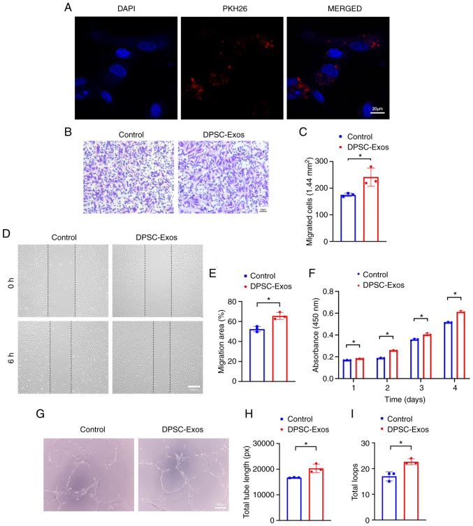
DPSC-Exos enhance the angiogenic activities of HUVECs. (A) Endocytosis of exosomes by HUVECs was visualized using fluorescent staining with PKH26. (B and C) The migration of HUVECs stimulated by DPSC-Exos was increased (n=3). Scale bar, 100 µm. (D and E) Representative images of scratch wound assay in HUVECs treated with DPSC-Exos or PBS. The remaining area of the DPSC-Exo-treated group was smaller than that of the control group (n=3). Scale bar, 100 µm. (F) The proliferation of HUVECs receiving different treatments was examined using CCK-8 assay. The DPSC-Exo-treated group exhibited a greater proliferative capacity than the control group (n=3). (G-I) Representative images of the tube formation assay on Matrigel in HUVECs treated with DPSC-Exos or PBS. The total tube length and quantity of total loops in DPSC-Exo-treated group were larger than those of the control group (n=3). Scale bar, 100 µm. *P<0.05. DPSC, dental pulp stem cell; Exo, exosome; HUVEC, human umbilical vein endothelial cell; PBS, phosphate-buffered saline.

